Is this a parasite?

luckykarma

New Member
I'm working with Appertex right now and hope to have great news within the next few days for the group.

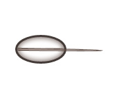
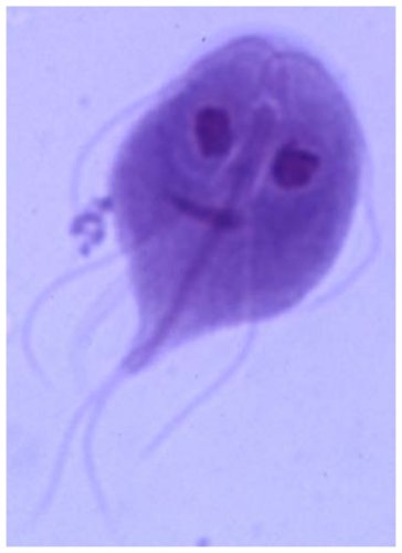
In the meantime as I do endless floats I see a good number of these things which I've drawn out in Photoshop. At first I thought it was a cricket part on top of a oil or small air bubbles but there was too many that were centered to be random.

These can be seen clearly at 400x. They're about the size of a parasite.
 

Attachments

  • parasite.jpg
    parasite.jpg
    3.1 KB · Views: 149
i believe that is a parasite just yesterday i took a stool sample of a pond and put it under the microscope and saw that same thing it had green circles in it as well and the antenna lookalike was the front of the parasite it used the antenna to navigate

i hope i helped

i was also wondering in order to see the chameleon fecal do you have to mix any liquids or anything to make the poop liquid so i can see through it ?

or do you take a little piece of it or what ?
 
No green anything and at 400x or 1000x it doesn't look like an antennae.

You have to use Fecasol and a fecalizer/slide/cover. If you want to do a smear you need saline solution.
 
i believe that is a parasite just yesterday i took a stool sample of a pond and put it under the microscope and saw that same thing it had green circles in it as well and the antenna lookalike was the front of the parasite it used the antenna to navigate

i hope i helped

i was also wondering in order to see the chameleon fecal do you have to mix any liquids or anything to make the poop liquid so i can see through it ?

or do you take a little piece of it or what ?

Based on Klingenberg Understanding reptile parasites 2nd ed:
Direct Smear:
smear the fecal matter to the slide
mix with few drops of distilled water

and cover the slide with cover slip.
you can use new methylene blue or Lugol's iodine to help bring out the contrast.
This is not exactly rewarding method.

Fecal float:
place small amount of feces in pill vial
fill vial with 2/3rd floatation solution (Fecasol, Ovasol, or saturated sugar solution) and break up the fecal material
add more solution until it reach the top of the vial
place cover slip on top the vial let it sit 10 minutes
take the cover slip, turn the wet side down on the microscope slide
examine for the parasite on microscope
 
To the best of my knowledge (I work for a veterinary parasitologist, although reptile parasites aren't my fortee...) this doesn't sound like a parasite. I think you very well could be seeing yeast or bacteria at 400X. Your description (without the magnification) could make it out to be giardia trophozoits ... but you would see those at 40x.

Really, 99% of parasites can be picked up at 10x and I never zoom in more than 40X.

Jamie
 
Thanks Jamie!

Dodolah gave the exact procedure for floating and smear. Sorry I didn't detail it out like he did.

However, my vet gave me a lecture about when I said I was using "distilled water" on the smear. His exact words were, "Why would you ever use distilled water?! No one on the planet would ever use distilled water for a smear!" and then went onto say the interaction etc.

Dr. Funk who gave the parasite lecture at the NABC last weekend also reiterated to use saline.
 
Saline should be more isotonic ... so if you're looking for a protozoan, you would maybe be more likely to find them intact. ALthough using saturated sugar isn't a good way to find protozoans, as the osmotic balance can be disrupted and destroy them. we recommend zinc sulfate solutions, but they can be hard to come by and difficult to dispose of properly.

Although for most non-protozoan parasites it really shouldn't matter.
 
Thanks Jamie!

Dodolah gave the exact procedure for floating and smear. Sorry I didn't detail it out like he did.

However, my vet gave me a lecture about when I said I was using "distilled water" on the smear. His exact words were, "Why would you ever use distilled water?! No one on the planet would ever use distilled water for a smear!" and then went onto say the interaction etc.

Dr. Funk who gave the parasite lecture at the NABC last weekend also reiterated to use saline.

lol.. ok saline solution it is :D
 
this is a stained giardia trophozoit. (yours not being stained would be clear instead of purple but the distinguishing features would still be present ... shape, eye like nuclei etc.). It doesn't look like this does it?
 

Attachments

  • stained giardia.jpg
    stained giardia.jpg
    128.8 KB · Views: 207
Jamie it doesn't look anything like that. What magnification is that photo? Do you know?

To my novice understanding smears are for protozoan parasites such as flagylites. Is that correct?

Would it be a good idea to use stain in smears?
 
Back
Top Bottom