JoeVet
Established Member
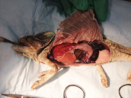
FWIW I am a veterinary Pathologist and I do like chams. I like the photos but I see a different story, closer to what Dr Ferret suggested. In the first picture, it looks like a filarid worm is on the left most rib, next to the bruise. Can't tell for sure without being there. Filarids are common in wild caught chameleons and can cause the rib bruising. I don't see fractured ribs but what I do see is total atrophy of the abdominal fat pad just below the cloaca (pic 4). My story would be that this is a parasitized animal in poor condition that died from stress and nutritional deficiency. He was not eating for the time you had him and the atrophied fat pad suggests he hasn't eaten for some time before you acquired him. We see one filarid worm which isn't likely the main cause of death but suggests there may be more unseen parasites and they may have contributed. A blood smear may pick up microfilaria in the blood if you have both male and female worms present. The lungs look good to me. It is congested but I don't see hemorrhage and the membranes and air sacs are clear. The one side is more congested due to blood settling in the down side (postmortem lividity). That's my guess. Next time give me a call and we can make slides.
Last edited: