xray-eggs??

idontspeekgeek

New Member
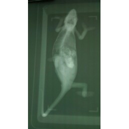
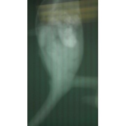
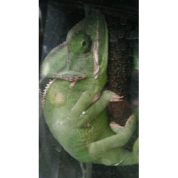
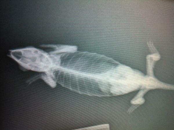
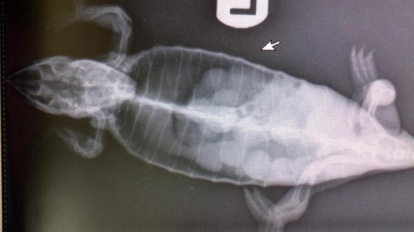
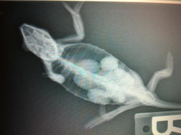
ok so i posted previously about i thought my chameleon had eggs in her. she still hasnt had them, and i took her to the vet this morning. he said if there are eggs in her, theyre very small still. can anyone else look at this and help me out. he also gave her a vitamin shot. shes recovering from mbd also.
 
looks like my pic didnt upload. ill try again just in case.
 

Attachments

  • koopaxray.jpg
    koopaxray.jpg
    6.8 KB · Views: 353
right click save to your computer. then zoom it. shows up well through a photo viewer.

The cloudiness up top doesnt appear to be eggs. but you can see a large amount of "ghost" egg outlines in her stomach.
 
i was sure there were eggs in her. im going to upload a couple other pics. thats the best one i have of the xray though. heres one for now.
 

Attachments

  • xrays.jpg
    xrays.jpg
    6.2 KB · Views: 286
in her back end below the white in the middle, you can see round things. and the vet said the white in the middle is her food?
 
Blown up shot. Large oval ghost structure on posterior half of body cavity might be maturing eggs? Tiny round eggs in mid section, would be eggs still in the ovary? Not sure what the large calcified area is in the middle- perhaps food in stomach?

femalevieledforum.jpg
 
in person i can see an actual cricket exoskeleton in her stomach. where you labeled around oval structure, i see that too. when i saw those, i could definitely see eggs in there. i was almost 100% sure those were eggs. calcifications?? im not sure if that is eggs or stomach...she ate 8 crickets yesterday and hasnt pooped. she doesnt have a problem pooping, she just hasnt gotten to it yet.
 
Blown up shot. Large oval ghost structure on posterior half of body cavity might be maturing eggs? Tiny round eggs in mid section, would be eggs still in the ovary? Not sure what the large calcified area is in the middle- perhaps food in stomach?

femalevieledforum.jpg

that is a good diagram gesang. i didnt even notice what you think are smaller uncalcified eggs. ohh geeze. i really hope that isnt the case.

and yes, i would tend to think the cloudiness in the middle would be food, seeing the solidity in it.
 
From that picture I do not see eggs. It would help to have another view from the side. The large most white structure (labeled "calcifications") is definitely stomach. You can't see follicles of the ovaries on a radiograph so those small roundish structures most toward the head is most likely just ingesta in the bowels mixing with gas. Everything else just looks like ingesta/fat pads. I don't see anything round or indicative of eggs. Doesn't mean they aren't in there hiding behind the other stuff, but I don't see any from that view. Did they take one from the side as well? That would help a lot.

I'll post some rads to compare.
 
***Don't be concerned about the tiny eggs. Those are the eggs in the ovary, ready to develop into the next clutch. Our veiled ladies often lay a secondary clutch about 60 days after a primary clutch. They'll do this about twice a year, for a total of 4 clutches in a 12 month period. There should be 2 ovary masses, each with tiny undeveloped eggs. And that what it appears to be in this case.

In the lower half of the body there appear to be eggs that have decended into the oviducts. From what I have been taught by our vets, there are 2 oviducts, one decending from each ovay (similar to human fallopian tube). The oviducts are very thin membrane "tubes", complete with their own bloodflow, from which the chameleon eventually lays her eggs, through the cloaca or vent opening.

*** I stand corrected - see ferret's comments quoted here below. Tiny egg-like stuff may just be bowel matter and gas. (Although I do want to say that I have an x-ray where follicular eggs were visible, we know because the vet showed them to us in the x-ray and then handed them to me after the girl had her ovaries removed. In that case the ovarian masses were huge, the follicular eggs were not being released, and so they had to be removed. So, it may have been an exceptional case.)


From that picture I do not see eggs. It would help to have another view from the side. The large most white structure (labeled "calcifications") is definitely stomach. You can't see follicles of the ovaries on a radiograph so those small roundish structures most toward the head is most likely just ingesta in the bowels mixing with gas. Everything else just looks like ingesta/fat pads. I don't see anything round or indicative of eggs. Doesn't mean they aren't in there hiding behind the other stuff, but I don't see any from that view. Did they take one from the side as well? That would help a lot.

I'll post some rads to compare.
 
Last edited:
These are early stage eggs:
full

See the distinct oval shapes?
K. multituberculata

This is one of the forum member's (sorry I can't remember who!) chams who had MBD and tons of eggs in very early development:
full

Veiled cham

This is eggs nearly ready to be laid:
full

Panther cham
 
Last edited by a moderator:
I had my female X-rayed several times with different angles each time and even a couple of ultra sounds and the vets could never be certain that she had eggs. This may not be your case but when a female is ill it seems that everyone often jumps to egg problems. I hope that all will work out for your girl.
 
Don't be concerned about the tiny eggs. Those are the eggs in the ovary, ready to develop into the next clutch.

No that is way too far cranial to be an ovary. Also you can't see the follicles on xray because it's all the same density and there's no contrast around it to give it shape - it's all the same density soft tissue. That consistency you're seeing is just food in the bowels. The darker area around the particles is gas (which is why you can see the particles). There is no gas in the ovary.

Chameleon Necropsy pictures - graphic
You can the ovaries and how far back they are on the first necropsy picture.
 
No that is way too far cranial to be an ovary. Also you can't see the follicles on xray because it's all the same density and there's no contrast around it to give it shape - it's all the same density soft tissue. That consistency you're seeing is just food in the bowels. The darker area around the particles is gas (which is why you can see the particles). There is no gas in the ovary.

Thank you for the explanation. I added a correction to my earlier post. But, just a question, if there is a clutch of larger eggs developing in the lower abdomen can it push the ovary forward? After taking another look at the xray I see 2 blurry light masses behind the "tiny white egg area". Those are prob the ovaries? Does the bowel appear distended or is that the normal circumference? Why is the bowel anterior to the stomach?


I had my female X-rayed several times with different angles each time and even a couple of ultra sounds and the vets could never be certain that she had eggs. This may not be your case but when a female is ill it seems that everyone often jumps to egg problems. I hope that all will work out for your girl.

Actually I think she's being treated by the vet for MBD and the egg questionis just a secondary issue.
 
Last edited:
Back
Top Bottom